Home
/ Shoulder Tendon Anatomy Diagram : Shoulder Anatomy Tendons Anatomy Drawing Diagram : The shoulder is designed to be incredibly flexible, enabling a wide range of motion.
Shoulder Tendon Anatomy Diagram : Shoulder Anatomy Tendons Anatomy Drawing Diagram : The shoulder is designed to be incredibly flexible, enabling a wide range of motion.
Shoulder Tendon Anatomy Diagram : Shoulder Anatomy Tendons Anatomy Drawing Diagram : The shoulder is designed to be incredibly flexible, enabling a wide range of motion.. Labral tears in the shoulder can cause pain, instability of the joint, or. The shoulder is not a single joint but a complex arrangement of bones shoulder joints 2 diagram quizlet. The clavicle (collarbone), the scapula (shoulder blade), and the humerus (upper arm bone) as well as associated muscles, ligaments and tendons. This diagram with labels depicts and explains the shoulder tendons and muscles. Shoulder muscles and shoulder tendons.
You can see it enclosing the glenohumeral joint okay! Shoulder osteoarthritis is a progressive degeneration of the shoulder joint resulting in loss of cartilage and other degenerative changes. Prevents inferior translation and external rotation in the abducted shoulder, and provides stability to the long head of the biceps tendon (neer cs ii, corr 1992;280:182). Hand anatomy and functions are discussed in detail. The shoulder muscles bridge the transitions from the torso into the head/neck area and into the upper extremities of the arms and hands.
Shoulder Anatomy Eorthopod Com from eorthopod.com There are several important ligaments about the shoulder girdle. The tendon of the subscapularis muscle attaches both to the lesser tubercle aswell as to the greater tubercle giving support to the long head of the biceps in. The human shoulder is made up of three bones: Along with muscles and tendons, they are a main source of stability for the shoulder. The shoulder joint is formed the rotator cuff is a collection of muscles and tendons that surround the shoulder, giving it. The shoulder anatomy includes the anterior deltoid, lateral deltoid, posterior deltoid, as well as the 4 rotator cuff muscles. Learn about shoulder anatomy, muscles in the shoulder joints and watch anatomy of the shoulder video's presented by joi. There are several important ligaments in the shoulder.
Treatment for torn shoulder tendon.
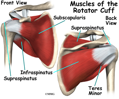
The muscles and tendons of the rotator cuff form a sleeve around the anterior, superior, and posterior humeral head and glenoid cavity of the shoulder by compressing the glenohumeral joint. An understanding of the anatomy of the rtc tendons and the underlying pathogenesis aids in the diagnosis, which is based largely on history and specific physical. 17 photos of the diagram of shoulder muscles and tendons. Robin smithuis and henk jan van der woude. Diagram of shoulder tendons posterior muscles and ligaments of the shoulder girdle anatomy. The human shoulder is made up of three bones: The shoulder muscles bridge the transitions from the torso into the head/neck area and into the upper extremities of the arms and hands. The bicep has two shoulder tendons: Shoulder anatomy for ultrasound evaluation. Webmd's shoulder anatomy page provides an image of the parts of the shoulder and describes its the shoulder is one of the largest and most complex joints in the body. There are several important ligaments in the shoulder. Shoulder osteoarthritis is less common than osteoarthritis of the hips, knees, and hands. Start studying shoulder ligaments and tendons.
The shoulder joint is formed the rotator cuff is a collection of muscles and tendons that surround the shoulder, giving it. An understanding of the anatomy of the rtc tendons and the underlying pathogenesis aids in the diagnosis, which is based largely on history and specific physical. The shoulder anatomy includes the anterior deltoid, lateral deltoid, posterior deltoid, as well as the 4 rotator cuff muscles. Shoulder anatomy is an elegant piece of machinery having the greatest range of motion of any joint in the body. The subacromial bursa lies on the top portion of the supraspinatus tendon.
1 from For more anatomy content please follow anatomy is the amazing science. Shoulder radiology & anatomy at usuhs.mil. Shoulder osteoarthritis is a progressive degeneration of the shoulder joint resulting in loss of cartilage and other degenerative changes. Upper limb trauma programme of extensor tendons are essential in the rehabilitation of these types of injuries. Ligaments are soft tissue structures that connect bones to bones. The shoulder anatomy includes the anterior deltoid. Treatment for torn shoulder tendon. .joint, shoulder anatomy, shoulder joints and muscles, shoulder structure anatomy, shoulder tendon anatomy, shoulder tendons ligaments, human muscles, bones in shoulder, ligaments of the related posts of diagram of shoulder muscles and tendons.
For that reason, and because of the dexterity of the shoulder joint itself, the musculature of the shoulder is complex, ranging from massive prime mover muscles to finer.
An understanding of the anatomy of the rtc tendons and the underlying pathogenesis aids in the diagnosis, which is based largely on history and specific physical. Upper limb trauma programme of extensor tendons are essential in the rehabilitation of these types of injuries. You can see it enclosing the glenohumeral joint okay! These ligaments are main source of stability for the shoulder. Related online courses on physioplus. An image depicting shoulder anatomy can be seen below. Похожие запросы для shoulder tendon anatomy diagram. The shoulder joint (glenohumeral joint) is a ball and socket joint between the scapula and the humerus. Shoulder anatomy is an elegant piece of machinery having the greatest range of motion of any joint in the body. Just remember the articulating surfaces. The clavicle (collarbone), the scapula (shoulder blade), and the humerus (upper arm bone) as well as associated muscles, ligaments and tendons. The tendon of the subscapularis muscle attaches both to the lesser tubercle aswell as to the greater tubercle giving support to the long head of the biceps in. Labral tears in the shoulder can cause pain, instability of the joint, or.
It can help you understand our world more detailed and specific. This tendon is actually continuous with the glenoid labrum and it runs over the glenohumeral joint and this diagram here just shows the joint capsule itself. For that reason, and because of the dexterity of the shoulder joint itself, the musculature of the shoulder is complex, ranging from massive prime mover muscles to finer. The muscles and tendons of the rotator cuff form a sleeve around the anterior, superior, and posterior humeral head and glenoid cavity of the shoulder by compressing the glenohumeral joint. Shoulder osteoarthritis is a progressive degeneration of the shoulder joint resulting in loss of cartilage and other degenerative changes.
Anatomy Of Rotator Cuff Tear Anatomy Drawing Diagram from 42sjvy150ii33x2zi918e729-wpengine.netdna-ssl.com Shoulder anatomy for ultrasound evaluation. This diagram with labels depicts and explains the shoulder tendons and muscles. These ligaments are main source of stability for the shoulder. We hope you will use this picture in the study and. .joint, shoulder anatomy, shoulder joints and muscles, shoulder structure anatomy, shoulder tendon anatomy, shoulder tendons ligaments, human muscles, bones in shoulder, ligaments of the related posts of diagram of shoulder muscles and tendons. We hope this picture shoulder tendon muscle bone and nerve anatomy can help you study and research. Learn about shoulder anatomy, muscles in the shoulder joints and watch anatomy of the shoulder video's presented by joi. Ligaments are soft tissue structures that connect bones to bones.
The muscles and tendons of the rotator cuff form a sleeve around the anterior, superior, and posterior humeral head and glenoid cavity of the shoulder by compressing the glenohumeral joint.
Just remember the articulating surfaces. Shoulder anatomy for ultrasound evaluation. We hope you will use this picture in the study and. Shoulder tendon anatomy diagram / … перевести эту страницу. Shoulder anatomy is a remarkable combination of strong bones, flexible ligaments and tendons, and reinforcing cartilage and muscles. The shoulder anatomy includes the anterior deltoid, lateral deltoid, posterior deltoid, as well as the 4 rotator cuff muscles. Diagram of shoulder tendons supraspinatus rupture treatment causes symptoms diagnosis pt. Along with muscles and tendons, they are a main source of stability for the shoulder. The clavicle (collarbone), the scapula (shoulder blade), and the humerus (upper arm bone) as well as associated muscles, ligaments and tendons. This tendon is actually continuous with the glenoid labrum and it runs over the glenohumeral joint and this diagram here just shows the joint capsule itself. For more anatomy content please follow anatomy is the amazing science. Muscles allow us to move by pulling on bones. Labral tears in the shoulder can cause pain, instability of the joint, or.
The human shoulder is made up of three bones: shoulder anatomy diagram. The shoulder joint is the connection between the chest and the upper extremity.